Category:Cyclic peptides
Jump to navigation
Jump to search
peptide chains which contain a circular sequence of bonds | |||||
| Upload media | |||||
| Instance of |
| ||||
|---|---|---|---|---|---|
| Subclass of | |||||
| |||||
Subcategories
This category has the following 12 subcategories, out of 12 total.
A
B
- Bottromycins (11 F)
C
- Ciclosporin (11 F)
E
- Echinomycin (6 F)
F
- Fungisporins (4 F)
M
- Microcystin-LR (7 F)
N
- Nodularin (4 F)
P
- Patellamides (5 F)
- Pentetreotide (3 F)
Media in category "Cyclic peptides"
The following 130 files are in this category, out of 130 total.
-
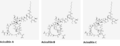 Aciculitin A-C.png 857 × 331; 12 KB
-
 ADH-1.svg 1,470 × 1,220; 30 KB
-
 Aerucyclamide A.svg 715 × 540; 24 KB
-
 Anidulafungin Molecular Structure.png 278 × 536; 17 KB
-
Artificial-human-Met-agonists-based-on-macrocycle-scaffolds-ncomms7373-s2.ogv 15 s, 960 × 540; 2.45 MB
-
 Bouvardin.svg 1,013 × 655; 49 KB
-
 Bremelanotide chemical structure.png 2,772 × 1,261; 18 KB
-
 Bremelanotide structure.svg 512 × 643; 19 KB
-
 Bremelanotide.svg 512 × 191; 20 KB
-
 Celogentin C.svg 954 × 359; 35 KB
-
 Chemical structure of Melanotan II Updated.svg 512 × 316; 9 KB
-
-
-
-
-
Cucurbitacin-I-Inhibits-Cell-Motility-by-Indirectly-Interfering-with-Actin-Dynamics-pone.0014039.s006.ogv 21 s, 1,114 × 460; 13.17 MB
-
-
 Cyclosomatostatin.svg 1,020 × 780; 52 KB
-
 Cyclotraxin B.svg 774 × 716; 137 KB
-
 Cyclotriprolyl Formula V1.svg 280 × 269; 10 KB
-
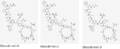 Discodermin B-D.png 785 × 318; 9 KB
-
 Discovery of Murepavadin.svg 8,078 × 4,413; 885 KB
-
 DPDPE structure.svg 1,755 × 1,385; 40 KB
-
Dual-Action-of-BPC194-A-Membrane-Active-Peptide-Killing-Bacterial-Cells-pone.0061541.s001.ogv 3.5 s, 589 × 444; 59 KB
-
Dual-Action-of-BPC194-A-Membrane-Active-Peptide-Killing-Bacterial-Cells-pone.0061541.s002.ogv 3.3 s, 589 × 444; 39 KB
-
Dual-Action-of-BPC194-A-Membrane-Active-Peptide-Killing-Bacterial-Cells-pone.0061541.s003.ogv 3.3 s, 589 × 444; 162 KB
-
 Enlicitide chloride.svg 512 × 365; 26 KB
-
 Enramycin strucutre.jpg 727 × 576; 64 KB
-
 Eptifibatide structure.svg 512 × 462; 16 KB
-
 Eptifibatide.svg 2,931 × 1,981; 54 KB
-
 Etamycin.png 360 × 295; 13 KB
-
 Fengycin.svg 751 × 958; 23 KB
-
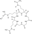 Ferrichrome - Ferricromo.png 2,357 × 2,537; 60 KB
-
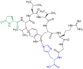
 Ferrichrome A.svg 1,190 × 805; 67 KB
-
 Ferrichrome.png 679 × 586; 102 KB
-
 Figure 3 (6998779227).png 752 × 760; 92 KB
-
 Friulimicin.svg 180 × 180; 16 KB
-
 Gene Cluster for Cyclothiazomycin Family.png 1,230 × 654; 62 KB
-
 Geodiamolide A.svg 447 × 316; 41 KB
-
 Geodiamolide B.svg 447 × 316; 43 KB
-
 Geodiamolide C.svg 447 × 316; 42 KB
-
 Geodiamolide D.svg 447 × 316; 39 KB
-
 Geodiamolide E.svg 447 × 316; 40 KB
-
 Geodiamolide F.svg 447 × 316; 39 KB
-
 Glidobactin A.svg 1,280 × 435; 24 KB
-
 Himastatin.svg 1,885 × 730; 78 KB
-
 Hymenistatin I.svg 990 × 830; 82 KB
-
 IRGD.svg 1,190 × 897; 66 KB
-
 Koranimine.svg 512 × 468; 24 KB
-
 KoranimineACS.png 692 × 633; 17 KB
-
 Lactocillin.svg 840 × 1,155; 125 KB
-
 Lanreotide.svg 1,320 × 1,100; 71 KB
-
 Lysocin E.svg 559 × 625; 35 KB
-
 Malacidin.svg 512 × 412; 26 KB
-
 Malacidin.tif 1,892 × 1,490; 259 KB
-
 Melanotan II.svg 512 × 631; 18 KB
-
 Merotocin.svg 2,550 × 2,080; 54 KB
-
 Microviridin B.png 1,620 × 2,044; 221 KB
-
 Moroidin V3(farblos).svg 715 × 588; 21 KB
-
 Moroidin.jpg 374 × 245; 16 KB
-
 Moroidin.svg 827 × 379; 39 KB
-
 Motixafortide.svg 1,830 × 1,345; 176 KB
-
 Murepavadin structure.svg 512 × 770; 92 KB
-
 Murepavadin.svg 2,557 × 4,288; 235 KB
-
 Mycobacillin.png 1,272 × 1,043; 36 KB
-
 Mycosubtilin.svg 2,607 × 2,207; 66 KB
-
 Neamphamide A.png 302 × 286; 5 KB
-
 Nepadutant.svg 2,565 × 1,560; 45 KB
-
 NIM811.png 724 × 686; 15 KB
-
 Nocathiacin I.png 498 × 353; 9 KB
-
 Nosiheptide.jpg 195 × 220; 13 KB
-
 Nosiheptide.svg 1,480 × 910; 60 KB
-
 Octapeptin chemical structure.png 358 × 218; 70 KB
-
 Octreotate.svg 2,652 × 2,622; 42 KB
-
 Octreotid V2(farblos).svg 629 × 458; 18 KB
-
 Octreotide.svg 2,652 × 2,622; 48 KB
-
 Pentixafor gallium-68.svg 1,815 × 855; 122 KB
-
 Pentixafor lutetium-177.svg 1,815 × 855; 120 KB
-
 Pentixafor.svg 1,815 × 855; 88 KB
-
 Pentixather lutetium-177.svg 1,815 × 855; 121 KB
-
 Pentixather yttrium-90.svg 1,815 × 855; 122 KB
-
 Pentixather.svg 1,815 × 855; 89 KB
-
 Peptide NR58-3-14-3.gif 288 × 354; 4 KB
-
 Perthamide C.svg 561 × 433; 27 KB
-
 Petrosifungin A.svg 605 × 745; 28 KB
-
 Petrosifungin B.svg 605 × 820; 29 KB
-
 Phakellistatin 13 Structure.svg 512 × 550; 250 KB
-
 Photobiotic 1.jpg 420 × 716; 58 KB
-
 Photobiotic.jpg 855 × 338; 57 KB
-
 PL-3994.svg 1,700 × 1,390; 95 KB
-
 Rencofilstat.svg 1,200 × 1,090; 53 KB
-
 Scyptolin A.png 250 × 297; 4 KB
-
 Setmelanotide.svg 2,270 × 1,720; 82 KB
-
 Structural formula of phakellistatin-13.png 232 × 232; 8 KB
-
 Structure of Lugdunin.svg 248 × 248; 39 KB
-
 Structure of surotomycin.png 744 × 1,317; 24 KB
-
 Stylisin 1 and 2.png 544 × 336; 9 KB
-
 Sulanemadlin.svg 2,560 × 1,107; 95 KB
-
 Surfactin.png 3,222 × 2,289; 54 KB
-
 Surotomycin.svg 103,040 × 57,680; 89 KB
-
 Syringolin A.svg 885 × 435; 23 KB
-
 Syringolin B.svg 885 × 435; 23 KB
-
 Tachyplesins.png 574 × 327; 8 KB
-
 Terlipressin.png 1,432 × 1,235; 37 KB
-
 Ternatin.png 266 × 286; 4 KB
-
 Ternatin.svg 222 × 202; 13 KB
-
 Theonellamide F-.png 288 × 231; 5 KB
-
 Trunkamide RiPP.png 1,136 × 1,613; 117 KB
-
 Tyrocidin A.png 2,044 × 2,037; 41 KB
-
 Tyrocidine 3D.png 3,000 × 2,400; 517 KB
-
 Tyrocidine-A.png 4,500 × 3,000; 1.02 MB
-
 Urodilatin - Compound Summary (CID 16133394).svg 569 × 540; 160 KB
-
 Urodilatin CDDANP-95-126.gif 361 × 479; 632 KB
-
 Urotensin II-related peptide.svg 1,110 × 860; 51 KB
-
 Valspodar.svg 2,867 × 1,487; 55 KB
-
 Vapreotide.svg 512 × 461; 72 KB
-
 Venturamide A.png 2,194 × 2,133; 221 KB
-
 Venturamide B.png 2,194 × 2,283; 238 KB
-
 VIR-576 secondary structure.png 435 × 570; 30 KB
-
 Viroidin.gif 281 × 233; 6 KB
-
 Viroisin V2 transparent.svg 635 × 493; 19 KB
-
 Viscumamide Structure.svg 685 × 695; 25 KB
-
 Viscumamide.png 228 × 198; 4 KB
-
 Voclosporin v.02.svg 716 × 548; 89 KB
-
 Voclosporin.svg 512 × 410; 17 KB
-
 Yunnanin A Structure.svg 512 × 388; 235 KB
-
 YUNNANIN A.png 295 × 220; 4 KB
-
 Zilucoplan structure.svg 512 × 331; 56 KB
-
 Zilucoplan.svg 4,473 × 840; 122 KB
-
 Zyklophin.svg 1,140 × 684; 75 KB


.png)


.svg)


.svg)


.svg)


